
Hot techniques in HD research
11 min read
Five HD scientists discuss the cutting-edge techniques driving their work. These technical innovations will provide biological insights and move us closer to treatments.
HD research is continuously evolving, as creative scientists develop and apply novel techniques to the study of cells, models, human tissue, and big data. In collaboration with the Huntington’s Disease Foundation (HDF), sponsor of the Huntington’s Disease Career Advancement Grant (HD-CAG), five researchers share here the cutting-edge techniques they’re bringing to their work, and why these innovations matter for accelerating discovery and moving the field closer to meaningful treatments. This article was jointly published in the Spring 2026 edition of HD Insights, a publication released by the Huntington Study Group.
How big is your repeat?
Carlos Chillon-Marinas and Sonia Vazquez-Sanchez, UCSD
Huntington’s disease (HD) is caused by an expansion of a short DNA sequence — the letters C-A-G — located in the very first part of the HTTHTT one abbreviation for the gene that causes Huntington’s disease. The same gene is also called HD and IT-15 gene. Everyone carries this repeat. The difference is how long it is. If the repeat is under 35 copies, people will typically do not develop HD. Above 40, the disease is fully penetrant.
The story does not end with the inherited mutation. Over time, this repeat can continue to expand within some vulnerable brain cells, a phenomenon known as somatic expansionsomatic expansion A process in which the CAG repeat in the Huntingtin gene can change over a person’s lifetime in some cells of the body, particularly in the brain.. This process is now a major focus of HD research, as it appears to be one of the key drivers of disease onset and progression. Ultra-long C-A-G expansions are strongly linked to cell damage.
This means HD is not only about the repeat a person inherits; it’s also about how much that repeat grows inside their brain. So how do you measure how these C-A-G’s expand?
Traditionally, repeat length has been measured using PCR-based techniques. These methods work by making a copy of the C-A-G repeat DNA and estimate repeat size from the resulting fragments that are made in the copying process. They work well for diagnosis and moderate expansions, but they struggle with very long repeats. Once repeats exceed roughly 100, the PCR-based methods become unreliable. The longest expansions — the ones that may matter most biologically — are often missed.
This is where PureTarget targeted long-read sequencing changes the game. PureTarget pulls out the entire repeat region molecule by molecule without breaking it into tiny fragments. These intact DNA molecules are then sequenced from end-to-end using high-fidelity long-read technology. This means that rather than guesstimating the CAG number, the researchers can actually read off the exact DNA letter code of the entire repeat region — along with the DNA surrounding it — in one continuous piece. Importantly, each molecule is sequenced multiple times, resulting in highly accurate repeat-length measurements.
For years, scientists suspected somatic expansionsomatic expansion A process in which the CAG repeat in the Huntingtin gene can change over a person’s lifetime in some cells of the body, particularly in the brain. was important, but our tools could not fully capture its scale.

The outcome is a much clearer view of repeat biology: exact repeat length, repeat interruptions, insertions or deletions, and variability between copies of the gene are measured directly rather than inferred. This matters because two cells with the same inherited mutation can accumulate very different repeat lengths over a lifetime, differences that may underlie selective neuronal vulnerability.
In a disease where DNA length drives biology, measuring the true size of the expansion is more than a detail. It’s a new window into how Huntington’s disease unfolds — and how we might one day slow it.
Multimodal spatial transcriptomics: looking at Huntington’s like never before
Sonia Vazquez-Sanchez and Carlos Chillon-Marinas, UCSD
One of the first questions scientists ask about Huntington’s disease (HD) is why the genetic change harms brain cells but spares other parts of the body, even though every cell carries the same CAG expansion. Even within the brain, not all neuronsneuron Brain cells that store and transmit information are affected equally. Some groups of neuronsneuron Brain cells that store and transmit information, such as those in the striatum, are remarkably vulnerable, while others remain resilient. For decades, researchers have been trying to understand why.
Traditional tools could only show fragments of the story. We could examine huntingtin proteinhuntingtin protein The protein produced by the HD gene. aggregatesaggregate Lumps of protein that form inside cells in Huntington’s disease and some other degenerative diseases, measure gene expression, or estimate the length of the CAG repeatCAG repeat The stretch of DNA at the beginning of the HD gene, which contains the sequence CAG repeated many times, and is abnormally long in people who will develop HD tract — but each experiment had to be done separately, often in many different samples of tissue. That meant we could only correlate results indirectly. We had no way to see all of these features at once inside the same cells, and we usually had to destroy the tissue’s natural structure in the process.
Multimodal spatial transcriptomics changes that.
This new approach is imaging-based and allows us to look inside individual cells while preserving intact tissue. It relies on advanced microscopy combined with a sophisticated fluidics system enabling the measurement of expression levels of thousands of genes, CAG repeatCAG repeat The stretch of DNA at the beginning of the HD gene, which contains the sequence CAG repeated many times, and is abnormally long in people who will develop HD length, huntingtin aggregatesaggregate Lumps of protein that form inside cells in Huntington’s disease and some other degenerative diseases, and signals of DNA damage, stress, and neuronal identity — all in the same experiment, and in the same cells, while maintaining their spatial context.
In essence, this technology lets us watch HD unfold with cell-by-cell resolution.
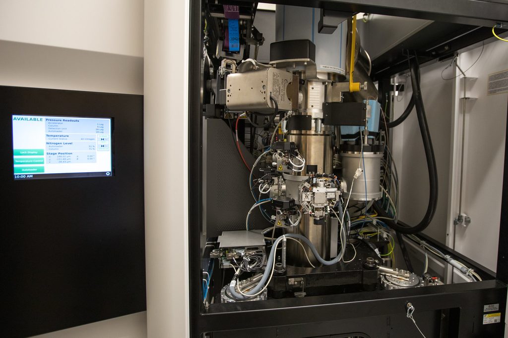
What makes this especially powerful is that it connects molecular events to what happens to individual neuronsneuron Brain cells that store and transmit information in HD. We can work out which cells are most vulnerable and begin to understand why: is it the size of the CAG expansion, the presence of protein aggregatesaggregate Lumps of protein that form inside cells in Huntington’s disease and some other degenerative diseases, or DNA damage? Early findings suggest that neuronsneuron Brain cells that store and transmit information with the largest repeat expansions are at greatest risk of loss, while huntingtin aggregatesaggregate Lumps of protein that form inside cells in Huntington’s disease and some other degenerative diseases appear more closely linked to shifts in gene activity. These relationships were previously invisible because no single method could capture all of these dimensions together.
Beyond solving long-standing biological puzzles, multimodal spatial transcriptomics may help guide future therapies. By revealing which DNA repair pathways are active in vulnerable neuronsneuron Brain cells that store and transmit information, or which cell types show the earliest signs of stress, researchers can make more informed decisions about therapeutic targets. As the technology continues to evolve, it promises an increasingly detailed view of how HD progresses within living neural circuits — and how we might intervene.
A high-resolution map of cells
Devon Pendlebury, University of California, Irvine
Cryo-electron tomography (cryo-ET) is an imaging technique which allows us to generate 3D pictures of important features inside cells. The cells are flash-frozen quickly, helping to preserve the insides of the cells to give us a more accurate picture of how the cells look in real life. Once frozen, an electron microscope is used to image inside the cells. Electron microscopes can see much more detail than traditional light microscopes. The frozen sample is tilted at multiple angles and a 2D image taken at each angle. These images can then be combined to create a 3D reconstruction which allows us to see compartments and proteins in the cell.
In HD, cryo-ET has been applied to see what aggregatesaggregate Lumps of protein that form inside cells in Huntington’s disease and some other degenerative diseases of the Huntingtin proteinhuntingtin protein The protein produced by the HD gene. look like in cells, and also to study how mitochondriamitochondria tiny machines inside our cells that process fuel into energy, enabling cells to function are impacted by mutant Huntingtin. A study by the laboratories of Wah Chiu (Stanford University) and Leslie Thompson (University of California, Irvine) utilized cryo-ET to look at mitochondriamitochondria tiny machines inside our cells that process fuel into energy, enabling cells to function, the energy factories of the cell, in HD neuronsneuron Brain cells that store and transmit information. They found that HD mitochondriamitochondria tiny machines inside our cells that process fuel into energy, enabling cells to function had distorted shapes and contained granules that were not found in healthy mitochondriamitochondria tiny machines inside our cells that process fuel into energy, enabling cells to function. This technique can also be used to determine how treatments affect mitochondriamitochondria tiny machines inside our cells that process fuel into energy, enabling cells to function health. The fine details available using cryo-ET allow scientists to identify changes in HD that would not be detectable through other methods.
The Force is strong with this protein: laser tweezers vs. Huntingtin
Piere Rodriguez-Aliaga, Stanford University
Do you remember Star Wars, Star Trek, or Ghostbusters, where light traps and manipulates ships and monsters? That technology was awarded the Nobel Prize in Physics in 2018. Optical tweezers are a single molecule “force microscope” that uses a tightly focused laser beam to trap tiny plastic beads. The beads act like handles: if a protein is attached between two beads using molecular linkers, the instrument can gently pull on that one protein molecule by moving the laser traps. While pulling, it measures both the force applied and how far the protein extends with nanometer precision. These forces are extremely small – typically a few to tens of piconewtons (billionths of a newton) – similar to what proteins experience inside cells.
This is particularly helpful for huntingtin because the region containing the polyglutaminePolyQ A description of HD and other diseases that are caused by abnormal expansion of stretches of DNA containing the sequence CAG repeated many times. Too many CAGs in a gene results in proteins with too many ‘glutamine’ building blocks, and glutamine is represented by the symbol Q. (polyQPolyQ A description of HD and other diseases that are caused by abnormal expansion of stretches of DNA containing the sequence CAG repeated many times. Too many CAGs in a gene results in proteins with too many ‘glutamine’ building blocks, and glutamine is represented by the symbol Q.) tract (the part of the protein that gets longer in HD) is wobbly, shifts between forms, and is prone to aggregation. Most structural methods work best when a protein is stable, uniform, and long-lived, but they report an average over huge populations of molecules. That averaging can blur distinct shapes together and hide rare, short-lived conformations that may still be biologically important – especially at polyQPolyQ A description of HD and other diseases that are caused by abnormal expansion of stretches of DNA containing the sequence CAG repeated many times. Too many CAGs in a gene results in proteins with too many ‘glutamine’ building blocks, and glutamine is represented by the symbol Q. lengths linked to Huntington’s disease.
Single-molecule optical tweezer experiments avoid this averaging by mechanically unfolding one huntingtin molecule at a time. By analyzing force–extension curves and unfolding/refolding event timing, researchers can map the protein’s energy landscape and conformations. They can determine whether the single protein (monomer) behaves like a random coil or forms a transient structure, whether intermediate structures exist, how stable they are, and how quickly the molecule switches between them. Because many individual molecules and measured, heterogeneity can be directly quantified rather than inferred from an average across millions of proteins.

Optical tweezers can help solve a long-standing question in the HD field: how does the genetic change render the huntingtin proteinhuntingtin protein The protein produced by the HD gene. toxic? Researchers are using this technique to compare huntingtin with different polyQPolyQ A description of HD and other diseases that are caused by abnormal expansion of stretches of DNA containing the sequence CAG repeated many times. Too many CAGs in a gene results in proteins with too many ‘glutamine’ building blocks, and glutamine is represented by the symbol Q. lengths and identify early structural changes that may trigger toxicity. They are also testing how post-translational modifications, chaperoneschaperone chaperone proteins help other proteins to fold correctly, and can protect proteins against damage, antibodies, and candidate drugs reshape huntingtin’s conformation. This single-molecule approach may reveal new therapeutic strategies, such as stabilizing a non-toxic huntingtin conformation. It may also provide mechanistic readouts for screening treatments and help connect molecular events to clinically relevant outcomes – showing how sci-fi technology can solve real biological problems.
Let’s sort it out
Chris Kay, Centre for Brain Research, University of Auckland
Your brain contains billions of specialized cells called neuronsneuron Brain cells that store and transmit information. There are hundreds or even thousands of types of neuronsneuron Brain cells that store and transmit information in the brain, each performing different functions, but strangely only a small subset die in HD. Scientists would like to know what makes these particular neuronsneuron Brain cells that store and transmit information so vulnerable to the disease, in the hope of learning how to stop them from dying in the first place. However, studying those vulnerable neuronsneuron Brain cells that store and transmit information is very difficult because they are surrounded by so many other kinds of unaffected cells in human brain tissue. If only we could sort out the vulnerable ones and examine them on their own!
This is exactly the idea behind a new technique being used by leading HD researchers to study what makes certain neuronsneuron Brain cells that store and transmit information different in HD, and why they are more susceptible to the disease. Fluorescence-activated nuclei sorting (or FANS for short) involves attaching a small fluorescent tag to a desired neuronneuron Brain cells that store and transmit information type in a brain tissue sample, then passing all the cells from that brain sample through a narrow tube with a camera. Whenever the camera detects a cell that is glowing with the fluorescent tag, it gets pushed into a separate tube for collection, whereas the non-glowing cells are allowed to pass straight through. After this process, the tagged neuronsneuron Brain cells that store and transmit information can be studied separately from all the other brain cell types.
Using these sorted neuronsneuron Brain cells that store and transmit information as starting material, scientists who study HD have already applied many cutting-edge technologies to investigate what makes them different. That’s what makes this novel FANS technique so powerful – once the neuronsneuron Brain cells that store and transmit information are sorted and separated, they can be analyzed in many different ways by researchers using a wide range of tools and methods. For example, some studies have already shown that the neuronsneuron Brain cells that store and transmit information most affected in HD have more somaticsomatic relating to the body CAG expansion. This is of interest to many drug companies that are trying to block somatic expansionsomatic expansion A process in which the CAG repeat in the Huntingtin gene can change over a person’s lifetime in some cells of the body, particularly in the brain. for the treatment of HD.
Takeaways
The future of HD research is driven by powerful new ways of looking at the disease. From reading DNA molecule by molecule to visualizing proteins and neuronsneuron Brain cells that store and transmit information in remarkable detail, these technologies are giving scientists an increasingly complete picture of how HD begins and progresses. With that clearer view comes a better chance of developing therapies that can make a real difference for those living with HD.
Summary
- Researchers are developing powerful new technologies that reveal HD biology in unprecedented detail.
- Long-read DNA sequencing can measure the full CAG repeatCAG repeat The stretch of DNA at the beginning of the HD gene, which contains the sequence CAG repeated many times, and is abnormally long in people who will develop HD expansion, helping scientists better understand somatic expansionsomatic expansion A process in which the CAG repeat in the Huntingtin gene can change over a person’s lifetime in some cells of the body, particularly in the brain. and how it contributes to disease progression.
- Multimodal spatial transcriptomics combines several measurements in the same cells, allowing researchers to connect repeat expansion, gene activity, huntingtin aggregatesaggregate Lumps of protein that form inside cells in Huntington’s disease and some other degenerative diseases, and cell health.
- Cryo-electron tomography creates high-resolution 3D images inside cells, revealing structural changes that cannot be seen with traditional light microscopy.
- Single-molecule optical tweezers allow researchers to study one huntingtin proteinhuntingtin protein The protein produced by the HD gene. at a time, uncovering fleeting structural changes that may trigger toxicity.
- Fluorescence-activated nuclei sorting (FANS) isolates the brain cells most affected by HD, making it possible to study them separately from surrounding cells.
- Each technique answers different questions, but together they provide a much more complete picture of how Huntington’s disease develops and progresses.
- These advances are accelerating basic research today and could help identify new biomarkers and therapeutic targets for tomorrow.
Sources & References
For more information about our disclosure policy see our FAQ…