
Are we finally getting that PET we’ve always wanted?
7 min read
For the past 20 years, PET tracers have been a game-changer for Alzheimer’s disease, making it possible to see amyloid plaques in the brain without invasive procedures. So what about Huntington’s?
Huntington’s disease researchers have been eagerly awaiting a new PET. No, not a puppy or a kitten or even a pet turtle, but a PET tracer to visualize huntingtin (HTTHTT one abbreviation for the gene that causes Huntington’s disease. The same gene is also called HD and IT-15) protein in living subjects. PET stands for Positron Emission Tomography, an imaging technique that can map protein concentration and distribution in the brains of living people. PET imaging uses a safe, injectable radioactive chemical, called a tracer or a ligand, which is designed to bind to a specific protein in the brain. PET tracers for amyloidamyloid The main protein that builds up in the brains of Alzheimer’s disease patients beta have been around for decades, and have been a crucial component of clinical research and treatment for Alzheimer’s disease.
The CHDI Foundation, a not-for-profit biomedical foundation specifically focused on treatments for Huntington’s disease (HD), has led the charge in developing a similar tracer for HD, designed to bind to mutant forms of the HTTHTT one abbreviation for the gene that causes Huntington’s disease. The same gene is also called HD and IT-15 protein. Two recent papers from CHDI, in collaboration with researchers from Belgium, evaluate the most promising PET ligand to date.
What exactly is a PET tracer and how does it work?
A PET tracer is a compound designed to bind to a specific target (in this case, the HTTHTT one abbreviation for the gene that causes Huntington’s disease. The same gene is also called HD and IT-15 protein) so that it can be visualized. The tracer is tagged with a radioactive isotope, which is an unstable version of a chemical element that releases radiation. Let’s think back to your high school chemistry class for a second (if not, skip ahead to the next section – we won’t judge).
During the process of radioactive decay, the isotope tracer releases positrons that quickly collide with electrons from atoms in the surrounding tissues. This collision destroys each of the particles and converts their combined mass into energy in the form of gamma rays. A PET scanner has a ring of detectors that capture the gamma rays, and then a computer algorithm converts the signal into a 3D image showing the precise location and abundance of the target protein in the brain. Very complicated sounding, but very cool.
Wait, isn’t radiation bad for you?

Well, that depends…all of us are exposed to low levels of radiation every day. These exposures include radiation from the environment, such as minerals present in the soil or water, or from activities such as flying in an airplane or getting a dental x-ray. These low levels of radiation are not harmful. Most countries have regulatory agencies that set safe exposure limits to protect their people, because it is known that high levels of radiation exposure can be very dangerous.
The radioactive tracers used in PET imaging are engineered to be very short-lived, so they don’t pose danger to the patient. The most common tracers use fluorine-18, which stays around long enough to create an image in the brain, but breaks down rapidly afterwards. The radiation from the tracer is almost entirely gone from the body within 24 hours. This is important to keep the patient and their loved ones safe.
Why do we need a PET tracer for Huntington’s disease?
Just as amyloidamyloid The main protein that builds up in the brains of Alzheimer’s disease patients PET tracers have revolutionized the clinical approach to treating and managing Alzheimer’s disease, we expect that a PET tracer for HTTHTT one abbreviation for the gene that causes Huntington’s disease. The same gene is also called HD and IT-15 will do the same for HD. Currently, there are ways to calculate the amount of HTTHTT one abbreviation for the gene that causes Huntington’s disease. The same gene is also called HD and IT-15 protein in bodily fluids, such as the cerebrospinal fluidCSF A clear fluid produced by the brain, which surrounds and supports the brain and spinal cord. that bathes the brain and plasma (a part of the blood), but there is no way to directly visualize the clumps of HTTHTT one abbreviation for the gene that causes Huntington’s disease. The same gene is also called HD and IT-15 protein, called “aggregatesaggregate Lumps of protein that form inside cells in Huntington’s disease and some other degenerative diseases,” in the living brain. HTTHTT one abbreviation for the gene that causes Huntington’s disease. The same gene is also called HD and IT-15 aggregatesaggregate Lumps of protein that form inside cells in Huntington’s disease and some other degenerative diseases build up in the brain as the disease progresses and are the target of many new drug treatments, so understanding how much there is and if a drug is changing that will help with developing medicine for HD.
A reliable PET tracer would show the specific location and intensity of the toxic HTTHTT one abbreviation for the gene that causes Huntington’s disease. The same gene is also called HD and IT-15 aggregatesaggregate Lumps of protein that form inside cells in Huntington’s disease and some other degenerative diseases in a living person. With so many new drug treatments designed to lower levels of the mutant HTTHTT one abbreviation for the gene that causes Huntington’s disease. The same gene is also called HD and IT-15 protein or prevent the formation of HTTHTT one abbreviation for the gene that causes Huntington’s disease. The same gene is also called HD and IT-15 aggregatesaggregate Lumps of protein that form inside cells in Huntington’s disease and some other degenerative diseases, PET imaging will allow a direct way to test how well these treatments are working in the brain and specifically where they are working. Further, it will allow doctors to select people who might be best suited to participate in a specific clinical trialclinical trial Very carefully planned experiments designed to answer specific questions about how a drug affects human beings, and will enable researchers to track how the HTTHTT one abbreviation for the gene that causes Huntington’s disease. The same gene is also called HD and IT-15 aggregatesaggregate Lumps of protein that form inside cells in Huntington’s disease and some other degenerative diseases match up with disease symptoms and outcomes.
“PET imaging will allow a direct way to test how well [HD] treatments are working in the brain and specifically where they are working.”
How close are we?
Over the past decade, CHDI has spearheaded a comprehensive program to tackle the challenges of developing a PET tracer for HTTHTT one abbreviation for the gene that causes Huntington’s disease. The same gene is also called HD and IT-15. HDBuzz previously covered this topic back in July of 2025, and we promised an update when we had more news – today is the day!
In that 2025 study, a PET tracer called CHDI-180R that had been extensively characterized in mice that model HD and monkeys was finally tested in live humans. Unfortunately, that tracer was deemed unsuitable for clinical studies, mainly due to lack of specificity for the toxic HTTHTT one abbreviation for the gene that causes Huntington’s disease. The same gene is also called HD and IT-15 protein and poor reproducibility when tested in the same person twice. Other versions of CHDI-180R were developed, but did not perform better than the original.
In late 2025, a new class of tracers was identified based on a different molecular structure. The performance of one of these new tracers, called CHDI-385, was the subject of two papers published earlier this year, in collaboration with the same group from Belgium.
What did the studies show?
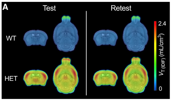
In the first study, the PET tracer (which uses fluorine-18) was tested in mice that model HD and showed increased specific binding to the toxic HTTHTT one abbreviation for the gene that causes Huntington’s disease. The same gene is also called HD and IT-15 protein compared to the previous tracers. That means it showed up only in the Huntington’s mouse brain and not in a control mouse that does not have the HD gene. The tracer also worked in young HD mice, who had very low levels of the toxic protein. This part of the study is relevant because a sensitive tracer could help identify individuals who are early in the disease process.
In addition, the new PET tracer appeared to be stable, was retained in the brain, but also could be cleared. This was similar to what they had observed with their initial tracers. These features are important to make sure that the tracer stays in the brain long enough to specifically bind to its target, while washing away from non-specific targets. However, this new tracer was the first one to be more consistent when they tested it in the same animal multiple times. The researchers showed that PET images from repeated testing in the same mouse were nearly identical, indicating excellent reproducibility, one feature that had eluded them in their previous study.
In the second study, the group tested how the new PET tracer was distributed throughout the body and how much radiation the body absorbed after injection. Using a software program designed to calculate optimal radiation doses to people, the researchers showed that their lead PET tracer could be given multiple times within a year and remain below the maximum threshold determined by regulatory agencies in the United States and Europe.
So, what’s next?
As mentioned above, a PET tracer for HTTHTT one abbreviation for the gene that causes Huntington’s disease. The same gene is also called HD and IT-15 will be a game-changer for HD clinical care. But getting that PET that we’ve been waiting for will ultimately depend on how well the new PET tracer works in human subjects.
All signs suggest that CDHI-385 is the most promising candidate identified to date, but the next step will be to test this tracer in a clinical study to see if it performs as well in humans. Researchers are cautious in being too optimistic, given the past disappointments, but we’re getting closer than ever on this front.
Summary
- Scientists are getting closer to a PET tracer that can detect toxic huntingtin (HTTHTT one abbreviation for the gene that causes Huntington’s disease. The same gene is also called HD and IT-15) protein in the living brain.
- Radiation exposure from PET tracers is low and short-lived, and early data suggest repeat use is safe.
- A working tracer would let researchers see where HTTHTT one abbreviation for the gene that causes Huntington’s disease. The same gene is also called HD and IT-15 aggregatesaggregate Lumps of protein that form inside cells in Huntington’s disease and some other degenerative diseases build up and track them over time.
- A new tracer (CHDI-385) shows strong specificity, sensitivity, and reproducibility in mouse studies.
- Earlier tracers were less effective than we hoped, so human testing will be the real determining factor for whether this one works.
- If successful, this tool could accelerate drug development and improve clinical trials in HD.
For more information about our disclosure policy see our FAQ…