2025 HDBuzz Prize: MR-“why” am I feeling this way? How MRI is helping us understand why people with HD may sometimes be unaware of their symptoms
We’re proud to announce Jenna Hanrahan as a 2025 HDBuzz Prize winner! People with HD may be unaware of their symptoms, not out of denial but due to real changes in the brain. MRI scans give new clues about what is going on.
People living with Huntington’s disease (HD) experience changes in their thinking, memory, and behaviours. This is anarea of HD that is widely studied and fairly well understood. However, sometimes people with HD don’t realize these changes are happening. This is actually a symptom known as anosognosia. This symptom isn’t the same as being in denial, where a person avoids reality while still being aware of it.
A recent study used MRI to explore why anosognosia happens in people with HD and what parts of the brain are involved. The findings help explain why people with HD don’t see or feel the symptoms that the people around them notice and why that is important for safety and understanding of the disease. Let’s get into what the researchers found, and what this means for the HD community.
Ignorance isn’t always bliss
Anosognosia can be mislabelled as stubbornness or laziness and dismissed as denial of a diagnosis. It can create major challenges for people with HD, their families, and caregivers. If a person does not realise they are sick, they may refuse or delay medical support, make unsafe decisions, or perhaps strain their relationships with family members and caregivers who are trying to help.
This study aimed to understand what is going on physically in the brain when this challenge presents itself. If these scientists can see physical changes that could be linked to anosognosia, they can help physicians, caregivers, and researchers better support people with
HD and understand why they might be unable to recognize their own symptoms. With this knowledge we could reduce the tendency to blame the impacted person and break the “stubbornness” stigma.
A photoshoot for your brain
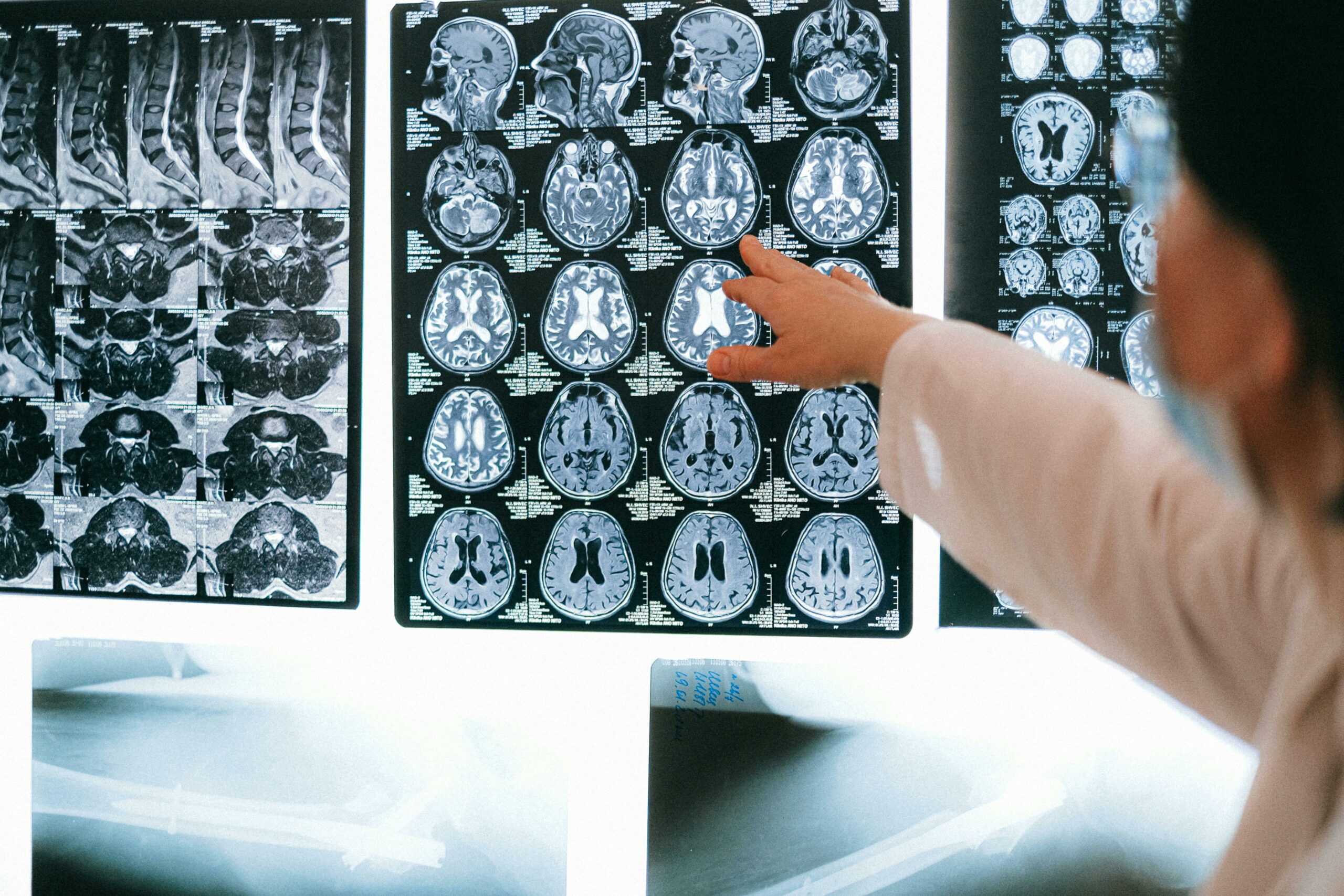
To investigate anosognosia, researchers looked at MRI (short for magnetic resonance imaging) scans from 570people (males and females). These scans came from two large research projects, the PREDICT-HD and TRACK-HD studies. Many participants were premanifest HD (HD-ISS stage 0), or in early stages of the disease (HD-ISS stage 1).
The MRI scans are essentially detailed pictures of the different structures inside the brain. Think of it like a super high-quality camera that uses magnets and radio waves to take pictures. Getting an MRI can be scary – it’s large, loud,and intimidating. But it is also powerful!

Here’s how it works: the machine is like the magnets on your fridge, except much larger and stronger. The hydrogen atoms that make up the water in our bodies act like micro magnets that are spinning in every direction (each one like a tiny Earth spinning on its own axis). When you enter the MRI machine, the big magnet makes the spins of the hydrogen atoms align. Then it sends out radio waves that push the hydrogen atoms out of place. Eventually they fall back to their original positions, giving off signals that the computer can detect. The computer takesall those signals and turns them into an image. All the thumping and buzzing sounds you hear are the quick electrical changes happening to the coils of the machine (the wires carrying the
electricity) to get the best images. It causes no damage, emits no radiation, and teaches us a lot about what’s happening inside our bodies – an all-round win!
But how do you measure a symptom someone is unaware of?
Since anosognosia means not being aware of your own symptoms, it can’t be measured directly. They used a questionnaire called the Frontal Systems Behaviour Scale (FrSBe). The person with HD and a companion who knows them well (caregiver or family member) filled out the survey. The questionnaire asked about things like behaviour, thinking and emotions. Comparing how the two responded to questions allowed them to observe the differences in how they view the symptoms (e.g., loss of energy, impulsivity, problems with organization).
Think of it like using two weather apps, where the app you use consistently misreports the temperature. If your weather app says it’s 20°C, and a friend’s app says 0°C for the same location, you may go outside without a sweater because you didn’t get an accurate reading of what the temperature really is. In the same way, a person with HD may think they are fine and rate their symptoms low, while a caregiver who sees what the symptoms truly are will rate their symptoms high. The bigger the gap in scores, the more unaware the person is of their symptoms, potentially impacting the person’s care.
The group used this gap to measure how much anosognosia the person with HD was experiencing. Then they ran the numbers, looking for connections between these score differences and saw is this tallied with what they saw in the MRI scans.

What did the study find?
The study found that seven brain regions were closely linked to anosognosia in people with HD – the globus pallidus, putamen, caudate, basal forebrain, substantia nigra, angular gyrus, and cingulate cortex. Each of these regionshas a special job, but many of them help with movement, learning, memory and emotion.
Through further investigation, one region in particular stood out: the globus pallidus. This region was thestrongest predictor of anosognosia in the study. This means that people with more globus pallidus atrophy (shrinkage) tended to be less aware of their symptoms.
The globus pallidus helps in controlling movements and makes sure our movements are smooth and efficient. In many HD studies and in the clinic, researchers tend to focus on the caudate and putamen, but this study suggests that changes to the globus pallidus could contribute to some of the symptoms of HD, especially when it comes to anosognosia.
What does this mean for HD families?
This research shows that anosognosia in HD is not about having a bad attitude, it’s linked to real brain changes. Being able to recognize the neurological side of things gives families and caregivers a better understanding of why people might not see their symptoms. It reduces the stigma and blame that is often misunderstood, and empowers safer decision making, and opens doors to new care strategies in the future. When we recognise that someone isn’t ignoring their illness on purpose, we can replace frustration with compassion.
Finally, a huge thank you to all the participants and families who contributed to the data sets used in this study. Your donation of brain data is a generous decision that will continue to provide new information and advance research. The studies you help build, like this one, stress the importance of working with the brain, and not against it and brings us one step closer to fully understanding the HD brain!
TL;DR
- People with HD often experience anosognosia (a lack of awareness of their own symptoms).
- A recent study used MRI data from over 500 people to explore brain regions linked to anosognosia.
- Researchers used a questionnaire to compare perspectives of caregivers and people with HD across different symptoms and used the gap between the two to measure anosognosia.
- Seven brain regions were linked to anosognosia, with the globus pallidus standing out as the most significant.
- These findings show that anosognosia in HD is linked to biological changes, not behavioral concerns. Knowing this can help families and caregivers respond with more empathy and better care.
Learn more
Meet this 2025 HDBuzz Writing Competition Winner

Jenna Hanrahan is a PhD student at Memorial University in Newfoundland, Canada, under the supervision of Dr. Lindsay Cahill. Her research focuses on using medical imaging techniques such as MRI and ultrasound to better understand brain changes associated with HD. Jenna hopes her findings will contribute to the development of novel therapies for the disease.
This year, the HDBuzz Prize is brought to you by the Hereditary Disease Foundation (HDF), who are sponsoring this year’s competition.

Sources & References
For more information about our disclosure policy see our FAQ…