Piecing It Back Together: Growing new neurons for Huntington’s disease
New research suggests the adult brain can be convinced to grow new neurons to replace those lost in Huntington’s disease. The SUPER exciting part? These cells don’t just grow—they seem to connect and function, like puzzle pieces clicking into place.

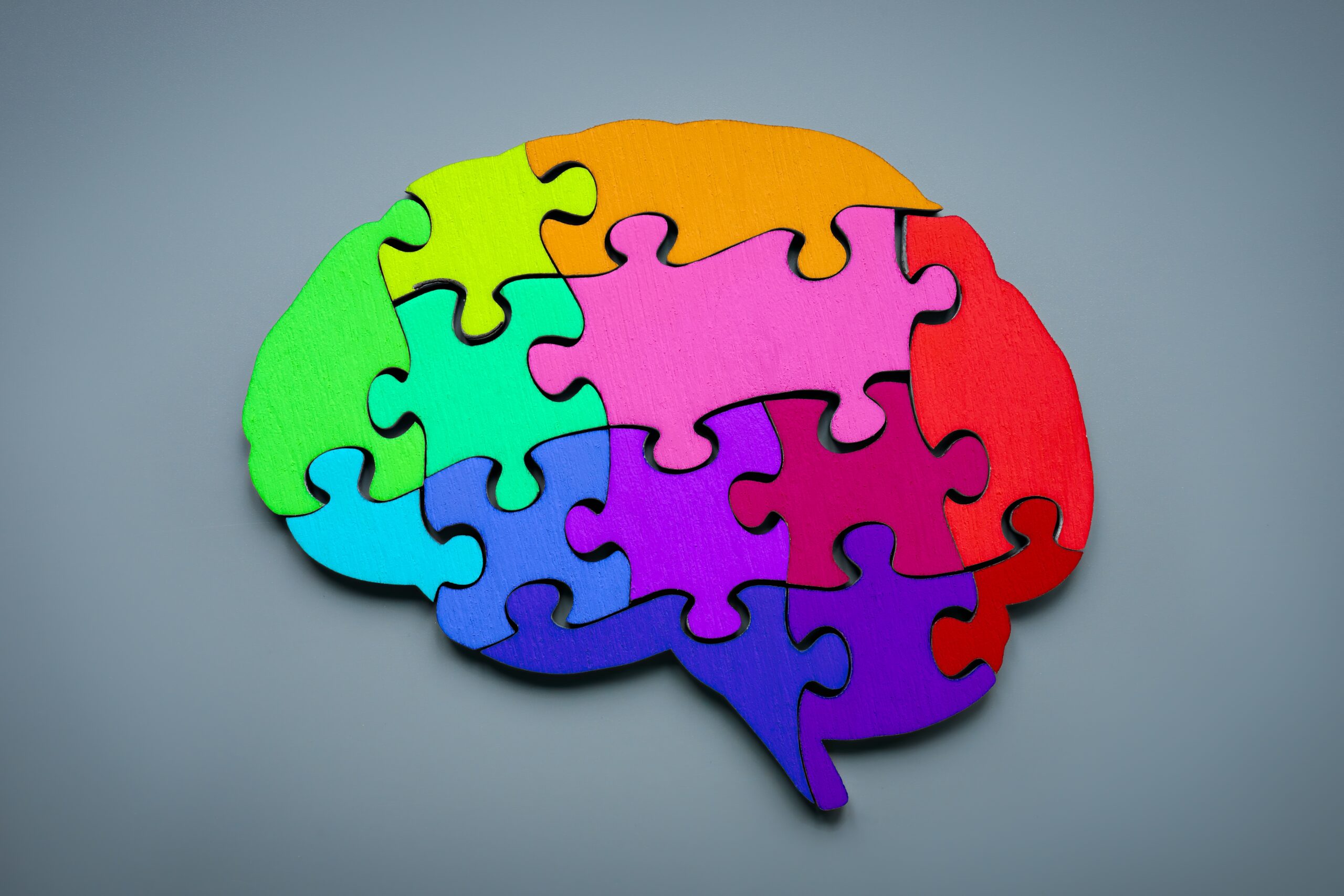
We often think of the adult brain like a completed jigsaw puzzle—once all the pieces are in place, that’s it. If a few pieces go missing, as happens in neurodegenerative diseases like Huntington’s disease (HD), there’s not much we can do except try to slow the loss. But new research is challenging that idea in a big way. A new study has shown that it may be possible to grow new brain cells in the adult brain—and not just any cells, but the exact pieces that HD takes away. Even more amazing? These new cells can connect with the brain’s existing networks, as if finding their place in the puzzle and clicking right into place. This discovery opens the door to a bold new goal: not just slowing the loss, but rebuilding the puzzle itself.
What Gets Lost in Huntington’s Disease?
To see why this study matters, let’s start with the pieces that go missing in HD. The disease causes progressive damage to an area of the brain called the striatum. The striatum sits almost exactly in the center of the head and helps control movement, emotions, and decision-making. The specific puzzle pieces lost here are called medium spiny neurons, or MSNs.

MSNs are essential connectors in the brain’s motor circuit. They help organize and relay instructions for smooth, coordinated movement. As HD progresses, these cells die off, breaking key links in the puzzle. The result: jerky movements, trouble thinking clearly, and emotional changes.
For a long time, scientists believed that once MSNs were lost, they were gone for good. But what if the brain has more pieces in the box—just waiting for the right signals to grow and fit into place?
Making New Pieces
That’s exactly what this new study from the lab of Dr. Steve Goldman at the University of Rochester set out to test. The researchers tried something bold: encouraging the adult mouse brain to grow new neurons. Not just any neurons, but the right kind—the ones that fit the MSN-shaped holes in the HD puzzle.
They used two special proteins to create the right environment. One, called BDNF (brain-derived neurotrophic factor), acts like fertilizer for neurons, helping them grow and survive. The other, called Noggin, guides stem cells toward becoming neurons rather than other cell types.
Think of BDNF and Noggin as puzzle guides: one boosts the brain’s ability to make new pieces, and the other makes sure those pieces are shaped correctly. When these were delivered to the brains of adult mice, something remarkable happened: the brain started creating new neurons that looked and acted like MSNs.
Lighting Up the New Pieces
To track these new cells, the researchers used a clever genetic trick that made newborn neurons glow under the microscope. This let them see exactly where new pieces were forming—and whether they matched the shapes of the ones lost to HD.
“A new study has shown that it may be possible to grow new brain cells in the adult brain—and not just any cells, but the exact pieces that HD takes away.”
In mice that received BDNF and Noggin, glowing new cells filled in the striatum. Many of them had the molecular markers scientists know are specific to MSNs. Even more encouragingly, they produced the same kinds of receptors MSNs use to communicate—essential for locking into the brain’s circuitry. It wasn’t just random growth. These were puzzle pieces that actually looked like they belonged.
Connecting the Dots
But pieces alone aren’t enough. For a puzzle to make sense, the connections between pieces matter just as much as the pieces themselves. The brain is no different. So, the next big question was: do these new MSNs actually connect with the right parts of the brain?
Using a safe, specialized version of the rabies virus (yes, really), the scientists traced incoming connections to the new neurons. They found that these newborn cells were receiving signals from all the right brain areas—the motor cortex, thalamus, and more.
Then, they flipped the direction and looked at where the new neurons were sending signals. Sure enough, they were linking up with the globus pallidus, a region of the brain that relies on MSN input to control movement. The puzzle wasn’t just getting new pieces—it was starting to fit back together.
Functional, Not Just Decorative
For the brain, even a puzzle with the right shapes won’t help if the pieces just sit there. To really matter, these new neurons had to be active and firing correctly. So the scientists used high-tech tools—like optogenetics and electrical recordings—to see if the new MSNs were actually sending signals and responding.
Image credit: Sharon Snider
The answer? Yes. These weren’t passive bystanders—they were live, working parts of the brain. They could receive and send electrical signals. They acted like mature MSNs. In other words, they functioned.
This is crucial. It means the new pieces didn’t just look right and fit—they helped complete the picture.
Moving the Needle on Symptoms
The final and most important test: could these new neurons make a difference in how the mice moved?
The researchers used a technique called chemogenetics to selectively activate the newborn MSNs. When they did, the mice that model HD—who typically move very little—became more active. Turning on these cells improved movement.
That’s a big deal. It’s like putting a few critical pieces back into a jigsaw puzzle and suddenly seeing the image take shape again. The effect wasn’t just cosmetic; it made a real difference in behavior.
A Piece to Remember
“The idea that the adult brain can grow new, functional MSNs—and integrate them into circuits that matter—is a seismic shift.”
While this work is incredibly exciting for HD families, it’s important to remember that these new cells have the same genetic makeup as the rest of the cells in the brain. So for someone with the gene for HD, that means the new cells will also have the gene for HD. Which means they’ll likely start to show signs and symptoms of the disease eventually too.
The good news is that the newly created MSNs would be developmentally “younger” than the MSNs initially created during brain development. Since HD has a delayed effect, we could expect that the same might be true in the new MSNs. In people, that delayed onset could provide the sort of time to improve health span and extend lifespan.
The less good news is that this means that this type of approach would very likely require multiple treatment rounds to compensate for the continued loss of the new brain cells. But, from where we stand right now, that would be a welcome problem!
Another positive piece of news to keep in mind is that these studies were done in adult mice using a pretty severe HD mouse model. This shows that even in more advanced cases, the right cell population may still be around and able to respond to treatment by BDNF and Noggin. While mice aren’t a one-to-one comparison for people, that’s great news!
What It Means for HD Families
This study doesn’t offer a treatment—not yet. But it completely reimagines what might be possible. For decades, scientists have worked to slow or stop the loss of brain cells in HD. Now, they’re asking: what if we could replace them?
The idea that the adult brain can grow new, functional MSNs—and integrate them into circuits that matter—is a seismic shift. It gives researchers a new strategy. And for people and families affected by HD, it offers something else: hope.
The puzzle isn’t finished. Pieces are still missing. But now we may have found a way to craft new ones—and click them into place. As always, we’ll be following this story closely at HDBuzz, keeping you updated as the picture comes more clearly into view.
Learn more
For more information about our disclosure policy see our FAQ…