Youthful competitors: young brain cells oust the old
Replacing cells with HD in the brain could be an effective treatment strategy. Recent work shows that glia injected into mouse brains take over and oust the older cells, but for a surprising reason – because of age, not HD!

When you lose something, an easy solution can be to just replace it. But what if the something you’ve lost are cells in the brain? Can they simply be replaced? Some researchers have been working toward this for Huntington’s disease (HD) by injecting new cells into the brains of animal models. A recent publication that has garnered a lot of press looked at the effects of replacing cells in the brains of mice that model HD – with surprising findings. The work draws attention to a less well-known type of cell and could inform future studies.
The brain’s supporting cast
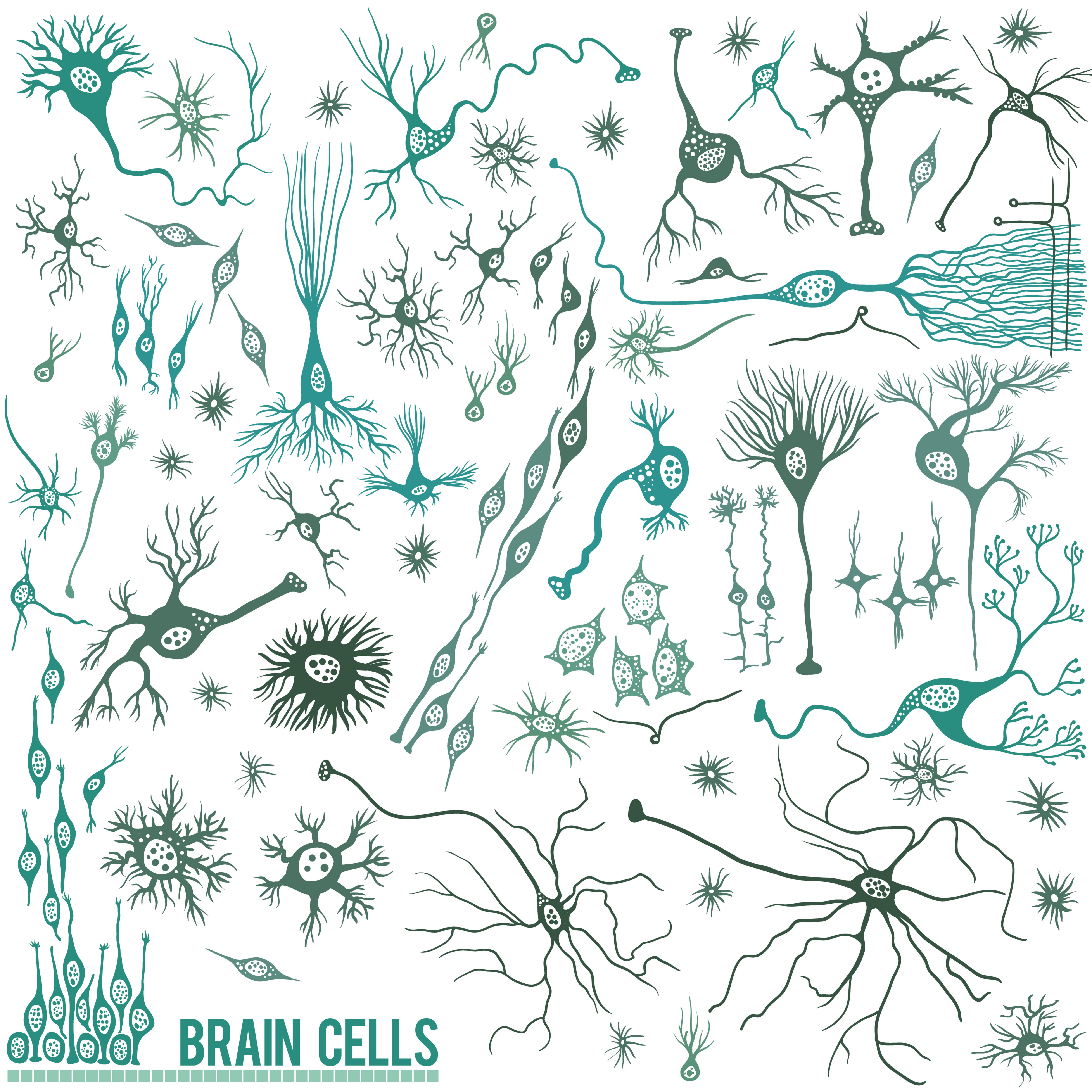
Neurons are one of many types of cells in the brain. They get a lot of attention in Huntington’s disease (HD), and rightfully so! Neurons are the cell type most affected by HD. They’re the ones that are shaped like a tree, with branches coming out the top, a long trunk, and roots at the bottom. This cell type transmits signals to help us think, feel, and move. We see neurons die over time in HD. But they’re not the only type of cell in the brain affected by HD.
Researchers are increasingly finding that other types of cells in the brain, called “glia”, contribute to HD. Glia are a support system for neurons in the brain, providing them with an environment that keeps them happy. We recently wrote about new findings related to the contribution of glia to HD.
Replace and improve
Back in 2016, researchers from New York and Copenhagen, Denmark did a series of experiments in which they replaced glia in the brains of mice that model HD. Excitingly, they showed that this improved the ability of the mice to function and delayed the onset of their HD-like symptoms. So even though glia aren’t the primary cell type affected by HD, replacing HD glia with healthy cells – ones that don’t carry the disease-causing mutation – led to a big improvement in mice that model HD!
Younger crowd taking over
Those same researchers, led by Dr. Steve Goldman, recently published follow up experiments to see if the same is true in human cells. But there was a twist – the experiments with human cells were done entirely in the brains of mice! They did this by creating a “chimera” – a single organism made from two genetically distinct populations. In this case, the brains of these mice had human glia containing the gene that causes HD.
The researchers wanted to know if they could replace the human HD-affected glia in the brains of mice by injecting unaffected glia. And they found that they could! When human glia without the mutation that causes HD were injected into the brain, they outcompeted the local human glia that had HD. The new healthy glia population took over, ousting the HD glia.
“So even though glia aren’t the primary cell type affected by HD, replacing HD glia with healthy cells – ones that don’t carry the disease-causing mutation – led to a big improvement in mice that model HD!”
Out with the old
But did the new glia take over in the mouse brain because they were healthy, while the resident glia had HD? Apparently not! The researchers also found the same results in the control counterparts for this experiment. In a surprise twist, the injected glia also replaced local glia that didn’t have HD. This suggests the replacement wasn’t related to the glia being sick with HD, but rather because the existing cells were older. The researchers found that the newly implanted glia were replacing the native glia simply because they were younger than the native cells.
The authors went on to perform molecular experiments to find out exactly what was going on. It turns out the new, young glia were just better at dividing, making it easier for them to take up space. Their presence also started a biological chain reaction that caused the older glia to die off. So it was really a one-two punch that allowed the young glia to outcompete the old – they were better at dividing, and they triggered the death of older glia.
What’s next?
The overall findings suggest that age was the primary factor for new glia taking over rather than HD itself. Even still, findings from this paper can help inform directions for HD research, particularly in relation to potential cell replacement therapies, like stem cell transplants.

Replacing lost cells could be beneficial for diseases like HD where we see a loss of brain cells that serve important roles in mood, movement, and behavior. However, we want to make sure the treatment itself doesn’t reduce the brain cell population that remains. In this publication, introducing new glia caused the widespread loss of native cells. While it may be good to have new glia, it could also be detrimental to lose glia that are already there.
Another point of caution for using this type of therapeutic approach for HD is that glia were replacing glia, not necessarily neurons. Since neurons are the primary cell type lost in HD, an effective treatment that replaces cells would also ideally increase the population of neurons in the brain. Future work should explore how a new and improved population of glia affects and influences neurons in the brain.
Researchers will also want to make sure that any treatment, whether it uses cell replacement or not, actually improves the symptoms of HD. Work described in this paper didn’t examine the behavior or overall health of the mice that model HD. So while they may have revamped their brains, we’re still not sure what, if any, effect this has on HD-like symptoms.
“The overall findings suggest that age was the primary factor for new glia taking over rather than HD itself.”
Overall, this paper brought us some cool science that shows that, in the case of human glia cell injections, cell replacement in the brain is possible. In the end, it was age that mattered more than disease. We’ll have to stay tuned to see if the fresh, young human glia improve the HD-like symptoms in mice, like the mouse glia did in the researchers’ 2016 paper.
For more information about our disclosure policy see our FAQ…